AMULET Innovality is a highly advanced mammography system with extremely fast image interval of just 15 seconds. This technology has ability to detect early breast cancer much more accurately than current mammography generations in Vietnam.
What is mammography?
Mammography is a technique that uses low-intensity X-ray beams to capture images of the mammary gland, thereby assisting radiologist to detect and analyze the presence of suspicious masses in the breast such as tumors, cysts and calcified areas...
This method helps to detect abnormalities and early stage cancers even when the patient has not yet felt and touched.
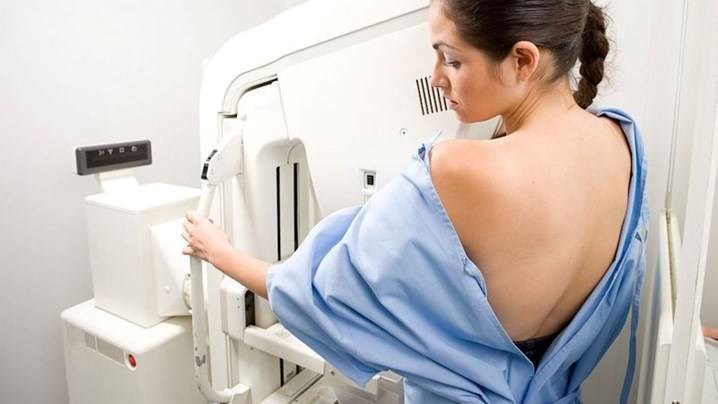
Photo: What is mammography?
Why need to take mammography?
In Vietnam, there are about 11,000 new cases of breast cancer annually and over 5,000 deaths each year. Mammography allows radiologists to detect abnormal lesions in the breast at an early stage, when treatment is easy.
Mammography are used to:
- Screening for breast cancer at an early stage
- Diagnosing breast disease.
When should I go for a mammograpphy?
You should go for a mammogram if you have one or more unusual symptoms, such as:
- Pain in the chest or mammary glands
- Abnormally large breasts
- A lump in the mammary gland
- Swollen armpit lymph nodes
- Changes in skin of the breast
- Nipple reduction, changes in the skin around the nipples
- Unusual bleeding or discharge
AMULET Innovality – digital mammography system at Hoang Long Clinic
The clinic was equipped with Fujifilm AMULET Innovality Mammography System, which captures 3D mammography for sharp images, short capture time, and higher accuracy than 2D mammography. This is the most modern and accurate mammography equipment currently available.
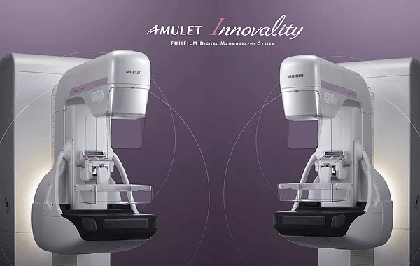
Photo: Fujifilm AMULET Innovality Mammography System in Hoang Long Clinic.
Tomosynthesis function for high quality 3D images enable superior diagnostic accuracy for easy diagnosis.
With tomography function, the x-ray tube moves in an arc to create a series of digital images at different angles and then these images are reconstructed into slices.
The reconstructed tomosynthesis slices makes it easier to identify lesions that are difficult to obtain with conventional mammography because of the overlap of other structures.
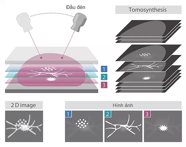
Photo: Tomography function help to observe the inner structure of mammary gland.
In this tomography function, ST mode combines rapid exposure timing and efficient workflow with a low x-ray dose, while HR mode makde it possible to to produce images with an even higher level of details, allowing the region of interest to be brought into clearer focus.
- HR mode (High Resolution)
- ST mode (Standard)
Iterative Super-resolution Reconstruction (ISR) method
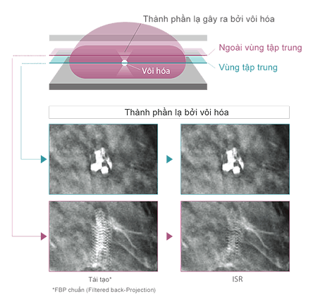
Iterative Super-resolution Reconstruction (ISR) integrated in Tomosynthesis was applied to optimize image quality and reduce X-ray dosage significantly.
- Reducing image artifacts in low-dose tomography
In this method, the images patterns are carefully recognized to selectively suppress the patterns that do not exist in human body architectures as noise, prevent artifacts in low-dose tomography.
- Suppressing interference of human body architectures at different depths (Reduce tissue overlap effect)
In the process of reconstructing the 3D breast architecture from multiple 2D images, calcification, mass, spicula, mammary gland and other signals that emerge from different depths in the breast architecture are selected off to reproduce the breast architecture at the focus depth with greater fidelity.
- Fine-structure restoration
Super resolution technology is introduced to restore the fine - structure of calcifications and other phenomena obscured by the x-ray tube movement, to facilitate interpretation of tomosynthesis images.
S-View – Synthesized 2D image
Tomosynthesis by AMULET Innovality automatically generates not only tomographic images obtained at 1mm intervals, but also a two- dimentional S-View images which is the combination of many 2D slices. showing the overwiew added to tomograms offering the views in detail, comprehensive image reading is possible.
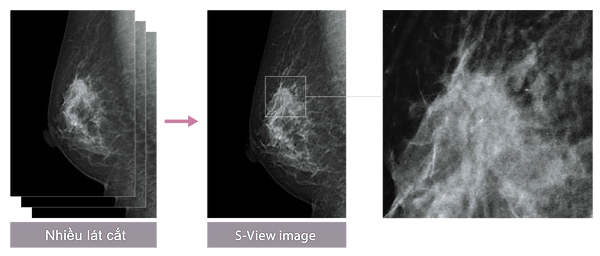
Photo: S-View – Synthesized 2D image
S-View images show the overview added to tomograms offering the views in detail, comprehensive image reading is possible.
- Gastrointestinal endoscopy - gold standard for early diagnosis gastrointestinal cancer
- Current methods of small bowel endoscopy
- AMULET Innovality – The most accurate mammography technology
- When to stop treatment for hepatitis B
- Endoscopic ultrasound – an advanced technique for diagnosing gastrointestinal tumors